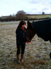
Today, my friends chiropractor came out to visit Spice as i was planning on riding her this weekend so just wanted to make sure that her back was o.k before i did but unfortunately she is a bit sore so i think i'll have to stick to just lunging her until the soreness subsides.
The chiropractor mentioned the possibility that it could be due to a condition called Polysaccharide storage myopathy (PSSM). However, it seems to be incredibly rare given the following article:
'A new test for a debilitating genetic muscular condition is now available in the UK and owners of horses predisposed to the condition are being urged to have them tested.
Polysaccharide storage myopathy (PSSM) is most prevalent in American draught breeds, paints, quarter horses and Appaloosas. Horses affected by the condition show signs of tying-up — muscle stiffness, sweating and a reluctance to move. PSSM is thought to be caused by an abnormally rapid uptake of glucose from the bloodstream.
Dr Stephanie Valberg of Minnesota University has carried out research into the condition. She told H&H: "I believe horses used for breeding or being considered for purchase, who show signs of muscle disease, should be tested for PSSM."
Until 2008, the only method of testing was via a muscle biopsy, but Minnesota University has developed a simple DNA test.
The research has also revealed that PSSM is passed on genetically via a dominant gene.
British Appaloosa Society chairman Brian Entwhistle said that PSSM is not yet an issue in the UK. He has been breeding Appaloosas for 40 years and has never had a horse with the condition. But he advocated testing for any genetic disease if a test is available.
"We don't want any disease being propagated within our gene pool," he said. "If there is any problem, I would expect all breeders to test their animals. I hope people will be open if there are diseases we should test."
Appaloosa breeder Alex McEachern said she has not experienced PSSM in 20 years of business, but wouldn't object to a buyer testing her horses.
But she added: "If it is a problem in America, might it not be a good idea to test breeding stallions before they can be licensed to breed in this country?"
To test for the disease send hair samples to the University of Minnesota Diagnostic Laboratory. Forms and contact information are available at http://www.vdl.umn.edu/vdl/ourservices/neuromuscular.html, or ask your vet to send a sample to the Royal Veterinary College in North Mymms, Herts. Testing of hair and root samples costs approximately £100 and a hair-only test is £43.'
So, i have to say i am a little doubtful of the prognosis but then again i haven't spent years training as a back specialist!
Below is another article i found on the condition:
Equine polysaccharide storage myopathy (EPSM) is a form of rhabdomyolysis classified as a metabolic disease that results in the accumulation of high muscle glycogen and abnormal polysaccharide in skeletal muscles.
The occurrence has presently been documented in Quarter horses, American Paint Horses, Quarterhorse crosses, warmbloods, draft horses and draft crosses. Presently, EPSM is believed to be transmitted as an autosomal recessive disorder with mares being more frequently diagnosed than geldings. Geldings may be more commonly affected with non-diagnosed clinical cases.
Horses that are affected generally are referred to as having a calm demeanor and being heavily muscled. Signs often occur 10-20 minutes after light work in 2-4 year olds starting training, but signs can also occur at any stage of life. The episodes may occur once or twice a year to every time the horse is exercised. A common complaint is that horses are exercise intolerant, especially at high speeds. In mild cases, horses show a tucked up abdomen, muscle fasiciculations in the flank, and a camped-out stance. If exercise is continued, profuse sweating, front and hindlimb gait asymmetry, and reluctance to move are seen. In severe cases, horses may refuse to move forward, buck and lie down to avoid exercise. When returned to the stall they may show signs of colic, such as rolling or pawing.
PSSM is a muscle disease in horses with Quarter Horse bloodlines such as Quarter Horses, American Paint Horses and Appaloosas. The American Quarter Horse Association (AQHA) has funded research into this disease since 1995 and has provided us with the opportunity to learn much about the diagnosis, cause and treatment for this disease.
Another form of polysaccharide storage myopathy also occurs in Draft, Draft crossbreeds, warmbloods. Many of the clinical signs in these breeds differ from those found in Quarter Horses and related breeds. The signs found in Draft, Draft crossbreeds, and warmbloods include muscle soreness, reluctance to engage the hind quarters, muscle atrophy, weakness, difficulty in backing up, and picking up hind feet.
Whether this disease exists in other breeds is controversial. Research from our laboratory suggests that there are several causes for tying-up. Most racing Quarter Horses, Thoroughbreds, Standardbreds and Arabians with tying-up, suffer from a separate disease from PSSM called recurrent exertional rhabdomyolysis (see RER information). In these breeds of horses, the accumulation of abnormal polysaccharide, the hallmark of PSSM, is rarely if ever present.
Several different acronyms have been used to describe this disorder including PSSM, EPSM and EPSSM. The variety of acronyms used are in part related to preferences of different laboratories, as well as to differences in the criteria used to diagnose polysaccharide storage myopathy. Many light breeds of horses, various draft breeds, ponies, warmblood breeds, and mules are reported to have PSSM when amylase-sensitive glycogen is used as a diagnostic criterion for PSSM. These criteria result in up to 80% of Draft horses and 33% of all horses outside of draft and Quarter Horse bloodlines being diagnosed with PSSM.
Polysaccharide storage myopathy (PSSM) is characterized by the abnormal accumulation of the normal form of sugar stored in muscle (glycogen) as well as an abnormal form of sugar (polysaccharide) in muscle tissue. About 200 horses of Quarter Horse and warmblood/draft horse breeding have been identified with tying-up associated with polysaccharide accumulation in muscles. This disorder is inherited in Quarter Horses and breeding individuals with PSSM has produced affected offspring.
Horses with PSSM accumulate muscle glycogen due to an unregulated uptake of sugar (glucose) into their muscles and the synthesis of its storage from in the muscle called glycogen. One aspect of the defect involves enhanced sensitivity of the muscles to insulin, resulting in more transport of sugar from the bloodstream to skeletal muscle. The diet can be adjusted to decrease the amount of insulin and sugar in the bloodstream. Carbohydrates that are high in starch, such as sweet feed, corn, wheat, oats, barley, and molasses, should be avoided and extra calories can be provided in the form of fat. An important part of the management of PSSM horses is daily exercise. This suppresses glucose uptake, enhances glucose utilization, and improves energy metabolism in skeletal muscle. If only the diet is changed, we found that approximately 50% of horses improve. If both diet and exercise are altered, then 90% of horses have had no or few episodes of tying-up.
An old theory about tying-up is that it is due to too much lactic acid in the muscle. Many exercise studies have proven that this is absolutely not the case with PSSM. PSSM is actually a glycogen storage disease and there are several diseases in other species and in human beings that also result in the storage of too much glycogen in skeletal muscle. In these other diseases, glycogen accumulates because the muscle lacks an enzyme (protein) necessary to burn glycogen as an energy source. These similarities led us to test PSSM horses for the disorders in glycogen metabolism identified in human beings. We found that PSSM is a unique glycogen storage disease because the PSSM horses have all the necessary enzymes to burn glycogen as a fuel in their muscles. With exercise, PSSM horses show the expected decrease in muscle glycogen as it is burned as fuel.
The unique feature of PSSM is that the muscle cells in PSSM horses remove sugar from the blood stream and transported into their muscle at a faster rate, and make more glycogen than normal horses. Our recent research shows that the reason for this is that PSSM muscles are very sensitive to insulin beginning as early as 6 months of age. Insulin is a hormone released by the pancreas into the bloodstream in response to a carbohydrate meal. It stimulates the muscle to take up sugar from the bloodstream. Once inside the cell the muscle’s of PSSM horses make much more glycogen than a normal horse.
The bits i have highlighted in bold are the symptoms Spice does NOT have so again i am not convinced that she has this condition.
But something is causing her to be sore (although two weeks ago according to another chiropractor she was only slightly sore around her withers?!?!)
The other thing about this condition that strikes me as odd is the fact that oil is suggested to be the only cure for it but where in the wild would a horse have access to a pint of oil or more each day?
As always i find two professionals in the horse world who seem to offer very different thoughts, opinions and advice so i have the usual dilemma of who to believe.
If i am completely honest i still think Spices hooves are at the root of the problem...below is an article which sheds some more light on this...
Low Heel/High Heel Syndrome
Unrecognized Problems
by Dr. Kerry J. Ridgway, DVM
The commonly observed condition where the heel of one front foot is higher than the other has ramifications that extend beyond the effects on the foot itself. This condition is also observed in the hind feet, though less frequently. However, because of limitations and scope, this paper will direct its attention primarily to the front feet.
How to best deal with the condition has remained a ‘hot topic’ among farriers. It is the aim of this paper to explore the often unrecognized ramifications in creating muscle imbalance, and changes in posture that result in loss of performance and are a potential source of lameness. It is necessary to explore how the syndrome alters the shape of the back and the consequences such changes create in being able to properly fit a saddle. The posture of the horse that results affects not only saddle placement but also alters the rider’s balance.
Awareness of the many issues came to me about 12 years ago via Moses Gonzales, journeyman farrier, when he demonstrated the effects that low heel/high heel syndrome had on the horse’s posture. Farriers and veterinarians, all too often, counter Gonzales’ observations with skepticism or antagonism. Healthy skepticism is always appropriate, so let us examine the issues on their merits.
In my career as a veterinarian specializing in muscle tension, imbalance and symmetry, I deal daily with performance issues, saddle related problems, shoeing related problems and back pain. These problems constitute as much as 90% of my practice. That has afforded me ample opportunity to observe the relationship of high heel/low heel conditions on a first hand basis. I believe that antagonism needs to be challenged and skepticism addressed. At the very least, this subject needs to be revisited with an open mind.
Let us first discuss the overall postural deviations that are a direct consequence of the lower of the two heels. It should be clarified at this point that it is not the intent to address a true ‘clubfoot’. This paper is not addressing an anatomically ‘short leg’ syndrome (though to a cursory evaluation, the limb with the lower heel may give the appearance of a shorter leg).
The lower heel creates obvious changes in the joint angles at the pastern, fetlock, elbow and scapula-humeral joint (shoulder joint). Compared to the limb with the higher heel, the angles on the low-heeled limb will open (get larger), and the limb will become more vertical than its counterpart throughout its length. The pastern joints and fetlock will be placed in more extension (and possible subluxation). The elbow angle will be more open. As the shoulder opens, the ‘point’ of the shoulder will be moved caudally so its position is farther back than on the higher heeled limb. The position of the scapula becomes altered so that it also becomes more vertical. This verticality creates a bulging of the shoulder and over-development of the associated muscles on the lower-heeled limb.
Observe that the horse has a marked tendency to lean on the shoulder of the lower heeled limb. This may leave some observers to conclude that the measurements that are to be described are ‘off’ only because the horse is leaning on that shoulder, and that if one pushes the horse to an equal weight bearing, the measurements tend to even up. However, this point must be addressed and clarified. We must answer why, given a choice, does the horse choose to lean on that shoulder? It is because of the difference in heel height that the horse returns to leaning on the shoulder of the low-heeled side when allowed to do so. I feel that this is the posture that the horse seeks as compensation.
Commence assessment of the forelimbs by observing the horse’s posture, its joint positions and angles from several directions. To have meaning, the horse must be on a flat surface. The horse must be standing ‘squared up’ on all four feet, and allowed to be bearing weight in its chosen posture. Ideally, the assessment is best performed after the horse has been trimmed, balanced, and is ready to shoe. Having one hoof placed even 3 to 4 inches ahead of or behind the other can alter the accuracy of the evaluation.
Start the observations from six to eight feet away in a position directly in front of the horse. Observe progressively from the foot upward, the position and relative heights of the joints. The foot with the lower heel will usually be significantly larger – the greater the size difference and the longer the low heel/high heel condition has been present, the more difference that will be evident. Difference in hoof size is a prime indicator that this condition exists. (While in this observation position also evaluates the coronary band for evidence of medial lateral balance.)
The fetlock joint on the lower heel side is generally lower than the higher heeled side. Next check the position of the styloid process of the radius. This is the ‘bump’ or ‘top of the shelf’ on the upper medial side of the knee (carpus0. It is nearly always lower on the low-heeled side. As the next step, evaluate and compare the height and symmetry of the points of the shoulder (scapulo-humeral joint). Generally it will be noted that the joint appears lower on the low heel side, and that there is hypertrophy of the descending pectorals on the side with the higher heel. With practice it can become evident that the shoulder point on the lower heeled side will also be place more rearward.
For an overall picture of the asymmetry, it can be very helpful to look at the spatial symmetry created by the inner margins of each limb and of the ventral aspect of the chest wall. In other words, look not at the limbs themselves, but use them as a “picture frame” of the space between the limbs.
Next, stand several feet away at the shoulder at a 90-degree angle from the horse’s direction of stance. From this position it is easy to see differences in pastern angles. In many instances, from this position, the pastern axis can be observed to be ‘broken backwards’. Broken pastern axis is accompanied by varying degrees of subluxation of the pastern joints. The toe may appear to be longer on the low heel side. The shoulder joint can now often be seen to be anywhere from 1⁄2 inch to 2 inches rearward of the limb with the higher heel. The difference in heel height is best seen by positioning one’s self another 45 degrees toward the rear of the horse and from about six to eight feet away.
Then the horse should be evaluated from behind and slightly above the croup. In order for a short person (or when examining a very large horse) to adequately make this evaluation, it helps to stand on a sturdy object of some sort. It will be noted that the shoulder of the limb with the lower heel will usually appear to have a significant lateral ‘bulge’ and it will appear to be higher than its counterpart. This is because the scapula has been displaced into a more vertical position. The shoulder with the higher heel will often appear to slope in an exaggerated manner.
Consider the consequences of this condition o n the fit of a saddle. The larger shoulder tends to exhibit some degree of muscle hypertrophy in the trapezius muscle. Other involved muscles may include the rhomboids, deltoids and subscapularis muscles. The trapezius muscle and the longissimus muscle support the fork or gullet bar in the fork, or head of the saddle. These muscles support the forward part of the bars or panels well. Saddles are for obvious reasons built symmetrically, so when placed on a horse with muscle hypertrophy on one side, the tree rotates diagonally into a position that allows similar contact on both sides of the ‘wither pocket’. Torque of the saddletree may make contact and place excessive pressure on one side of the thoracic spines and leave more openness on the opposing side. The result is pain, loss of ability perform bending and lateral movements. The pressure can also create chiropractic subluxation of the withers. The bulged shoulder may strike the edge of the panel or bar as the scapula moves through its range of motion.
Because the opposite shoulder typically has more slope, the saddle may tend to fall or slip to the sloping shoulder side of the horse. This is a second reason for pressure on the thoracic spinous processes. The problem of slipping to the side is particularly troubling if the croup is also involved and is lower on the same side. This can occur when a high/low condition exists in the hind feet as well. More often, however, when the horse is observed in motion, one side of the croup rises more on one side that the other. It relates to muscle balance, chiropractic or joint issues in the hind limbs. Regardless of cause, it creates an even worse scenario for slippage when combined with shoulder asymmetry.
A rider who must alter his/her position and posture because of improper position of the saddle, will eventually create performance problems and increase the risk of lameness. Most riders have allowed their bodies to compensate and are usually quite unaware of their compensation until it is brought to their attention. Additionally the rider may end up with chronic back, hip or knee pain. The horse, because of a ‘crooked rider’, will experience performance problems and eventual lameness. The crooked saddle and side slipping saddle causes the rider to place more weight in one stirrup than the other. This creates a ‘crooked’ traveling horse and is a cause of sub- clinical and eventually clinical lameness. There are many other postural deviations of the rider that can add to the problem.
It has been a consistent observation that horses that exhibit the high/low syndrome are frequently found to have chiropractic issues and muscle pain and spasm at the base of the neck. Chiropractic subluxations are present, more often (but not always) on the limb with the higher heel. As previously stated, the pressure a ‘crooked’ saddle places on one side of the thoracic spinous processes leads to pain and chiropractic subluxations of the upper thoracic vertebrae.
The consequence of such subluxations is one of the most common causes for a horse to react badly to the tightening of the cinch or girth. The subluxations create
neuromuscular irritability in muscles of the shoulder and in the area covered by the girth or cinch. Uneven weight bearing created both by the syndrome and the change in saddle fit frequently leads to suspensory and check ligament problems. Horses thus affected may also have trouble with a lead or lead changes and may tend to cross canter.
With regard to the foot itself, the syndrome produces a long toe with the heel becoming under-run. This, as we know from Dr. Robert Bowker’s work, leads to inadequate support in the posterior part of the foot and eventually to degeneration of the digital cushion. Digital cushion failure, when present, leads to a ‘broken pastern axis’ that is very difficult if not impossible to correct. It is not uncommon to see large, flat and splayed out frogs accompanying the foot with a degenerated digital cushion, as the front tries to compensate and support the posterior portion of the foot. There is an obvious consequence to be recognized with regard to factors creating ‘navicular disease.’
For the many reasons presented, I feel quite strongly that it is inadequate to address the foot without looking at the consequences on the topside of the horse. Without seeking and correcting the root cause (in this case the high/low heel syndrome) any other treatment is only palliative. By properly addressing the high heel, low heel syndrome, the farrier can be of enormous help to both the rider and the horse.
There have been many theories advanced as the reasons for the low heel. Regardless of the originating factor, whether genetic, or acquired, we are all aware that the horse will typically graze with the limb having the low heel advanced. It is certainly a reasonable theory that pressure (on the heel), maintained through many grazing periods, distorts the hoof capsule, unbalances the foot, advances the break- over location, and causes the heel to become under-run. Pressure over time creates distortion. Distortion equals an unbalanced foot.
What is the appropriate shoeing for this condition? I feel that a cardinal rule is to work primarily with the foot that has the lower heel. Additional problems are incurred if the heel of a true “clubbed foot” is lowered excessively. Structures in the muscles called “spindle cell receptors” and receptors in the musculo-tendonous portion called “Golgi bodies” provide signals from the muscle or tendon to the spinal cord. This data provides information to the central nervous system (CNS) about the tension that exists in the muscles and tendonous structures.
When the heel is lowered, the receptors in the deep flexor tendon are activated and signal the CNS that there is too much stretch in the tendon. The response from the CNS is to issue a signal to shorten the muscle or tendon structures to prevent injury. This response provides one reason why, at the end of a shoeing period, a clubfoot that has had the heel lowered, usually looks as bad or worse as when originally seen.
My experience has lead to the conclusion that the best course of corrective shoeing is the use of wedges as orthotic devices, applied on the lower heel in order to achieve the same heel height and pastern angel as the more upright foot. Sometimes it is necessary to also use a ‘lift’ such as a rim (or full) pad on the same or opposite foot as well, in order to create full symmetry. The determination of height of the wedge at the heel, or if a lift in one side or the other is required, is best determined after the horse has been trimmed and balanced. Again, it is emphasized
that this determination must be made with the horse standing squarely on a firm level surface.
Have the horse stand on the trial orthotics and re-examine for an improvement in symmetry. Use the same examination process as previously described. In review, check factors such as the symmetry of the space between the legs, height and angle of the joints, and the height of the styloid processes. Note whether the ‘point’ of the shoulder now comes into symmetry with the opposite side. Again observe from above and behind to determine the effect on angle and symmetry of the two scapulae. Sometimes the changes observed by this procedure are dramatic. In longstanding cases (especially in older horses) the changes are subtle, and immediate results are not as evident. But you will see improvement in symmetry and performance over the course of multiple shoeings.
Without proper attention to break-over and heel support, the overall condition of the foot can be made worse with this use of wedges. How can this be ameliorated? When shoeing this type of foot, the breaker-over must be set significantly back (to approximately 6 mm ahead of the tip of the coffin bone. A slightly exaggerated heel support should be used and at the very least should extend to the widest part of the buttress of the heel. The wedge should then extend roughly 1/8 inch beyond the heel of the shoe. Following these precautions helps to prevent the heel from further crushing. If the walls are already rolling under at the heels it is necessary to trim them lower yet, to the level where there is sound wall growing in the proper direction and not rolling under. It follows that radical trimming must be followed by using a larger wedge in order to create the appropriate heel height and pastern angle. For most cases, the author prefers an ‘open’ bar wedge, and where indicated, rim pads instead of full pads.
If there is evidence of digital cushion deterioration, it may be necessary to use full pads and impression material, as well as a frog support. In younger horses there is a better rate of success in re-directing the hoof wall growth, re-balancing muscle development, and maintaining a back that can appropriately accommodate a saddle. Quite a few of these horse may be taken out of all support after a limited number of shoeings, Older horses with very long standing problems and poor quality digital cushions are often best kept in the appropriate amount of orthotic on an ongoing basis.
Options included using a wedge shoe on the low heel side, wedging the heel, or simply leaving more heel on the low side. In our experience, the latter choice is the least desirable because the frog often loses the contact it needs to assist in any possible restoration of the digital cushion. If chosen, it should be accompanied by impression material and a frog support on the solar surface.
For muscle re-balancing to occur following postural correction, the horse must be in work. There is always concern with how much work is acceptable and how soon should work resume after shoeing corrections have been done. It is, of course, not a bad idea to err on the side of conservatism and work lightly for the first week or ten days. However, I have seen horses remain in athletic competition immediately after the shoeing changes. Even with no decrease in intensity or schedule of competition they have suffered no apparent negative effects.
One last point – if one is not happy with the results, it is a simple matter to remove the orthotics at any time. However, I believe that if the farrier considers all
the factors and shoes appropriately according to the principles discussed, the results will be positive for all concerned.
Friday, 28 May 2010
Subscribe to:
Post Comments (Atom)
No comments:
Post a Comment